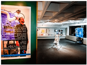
Dissertation: Biologically encoded augmented reality
This is a stick figure. The world we perceive around us is an incomplete image, of not
This is a stick figure. The world we perceive around us is an incomplete image, of not
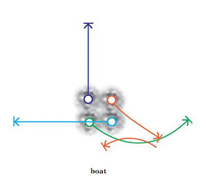
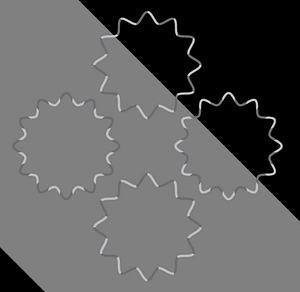
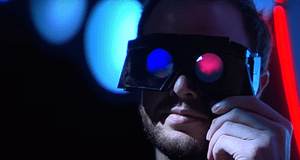
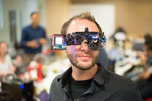
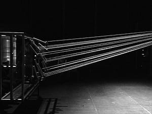
This research demonstrates a foundational approach to peripheral semantic information delivery capable of conveying highly complex symbols well beyond the established mean, using motion-modulated stimuli within a series of small, static apertures in far periphery ( > 50°).
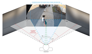
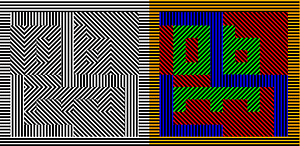
How detectable are far peripheral semantic cues in increasingly dynamic environments? Results show rapid adaptation and high detection accuracy.
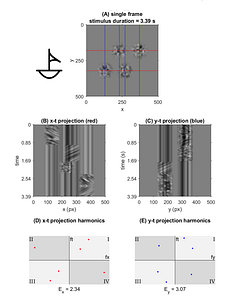
Metrics of symbol recognition speed and accuracy in a controlled environment, with codex parameterization using motion energy analysis
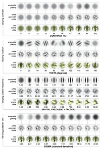
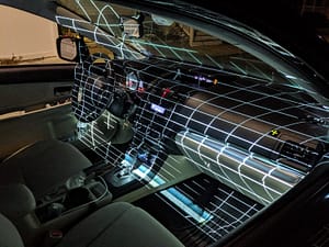
Adapting stimuli to dynamic environments One of the greatest challenges of adapting motion-modulated semantics to real-world environments
Information floods the center of our visual field and often saturates the focus of our attention, yet
The advancement of exploratory mediums to form new modes of representation functions as one of the many
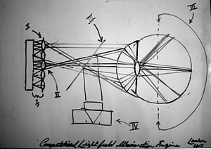
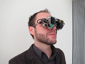
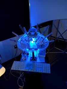
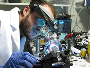
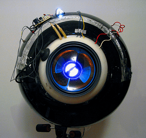
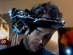
This system introduces ophthalmic light field capture, on a micro camera platform, allowing for a robust approach to the acquisition of multi-view imaging and ensuring nearly all light emitting from the pupil is gathered for processing.
The development of bionic and cybernetic systems illuminates new potentials for the future of non-invasive, networked, neuro-muscular
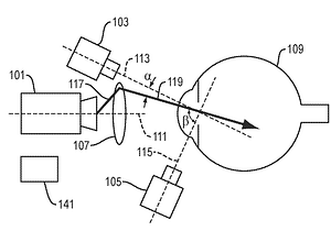
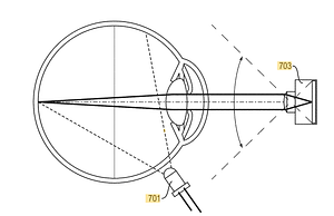
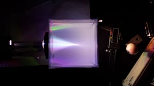
ABSTRACT: A projector and one or more optical components project a light pattern that scans at least
Our visual realities are flooded with complex and robust natural scenes. New findings in curvature estimation illustrate
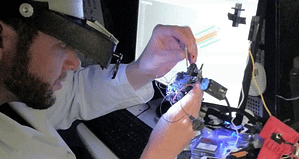
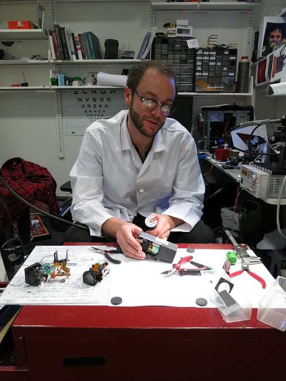
We are surrounded by displays and technologies whose mechanisms are hidden from view. This work was an
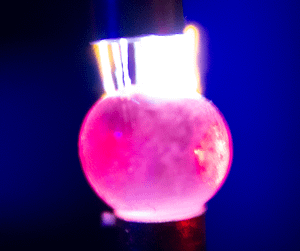
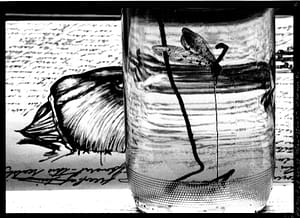
Spherical tissue deformation in electrostatic fields (NaCL+H20 suspension in hydrogel structure)
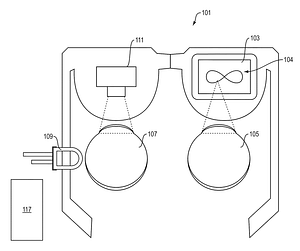
Neurovascular imaging interface, implementing coincident imaging/illumination path and through-pupil illumination.
Biomimetic systems architecture engineering: prototypes Quantizing visual perception as an expression of experience is referentially viewed as
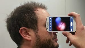
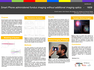
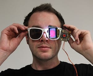
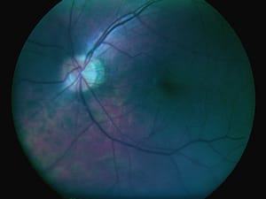
This self-directed, interactive device captures and visualizes images of the retina without the need for complex optics and precise alignment of illumination pathways.
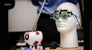
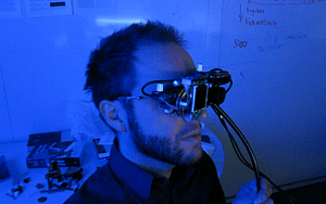
The eye is a highly sensitive and programmable array of sensors. By presenting stimuli to discrete regions

The following sequence presents a demonstration of the interlinkages of visual and auditory sensory processing. The video
Indirect, diffuse illumination may be employed, rather than direct illumination of the retina through the pupil. For
Angle, inertia, and velocity are a subset of moment arms that when instinctively activated are biologically hardcoded
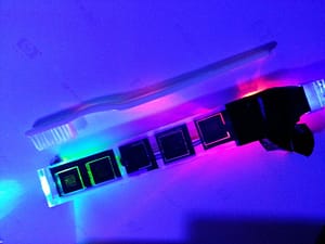
This research investigates the viability of subsurface imaging of the teeth and gums as a possible metric
Publications pending Please log in to view protected content:
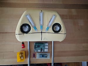
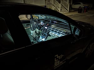
Proprioceptive feedback systems for kinetosis (motion sickness) treatment and prevention Rather than treating the environment, the automobile
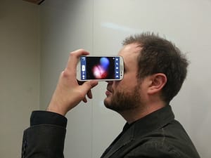
Non-mydriatic fundus imaging on smart phone platforms may prove to be a paradigm shift in rapid screening devices with no operational expertise required
This system expands upon prior work by fully integrating the self-alignment architecture within the portable user interface.
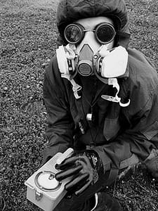
I confront the filters of cognition as an intervention into the control I assume to have over
A cataract-affected eye scatters and refracts light before it reaches the retina, caused by a fogging or
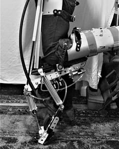
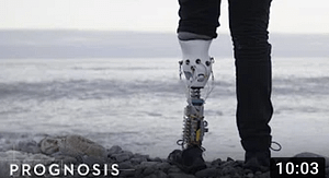
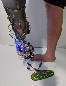
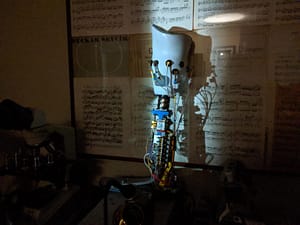
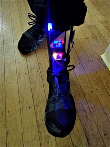
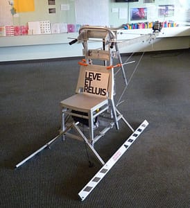
Power lift prototype: compressor force-resistive foot feedback with on-board mulit-channel solenoid valve configuration [lift capacity = 600
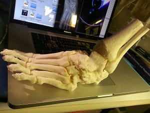
The pain I had been experiencing for months following my reconstructive ankle surgery was not identifiable by
A low-cost, wearable solid-state device with no moving optical parts is articulated to create a full, 3D reconstruction of the anterior segment of the eye.
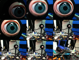
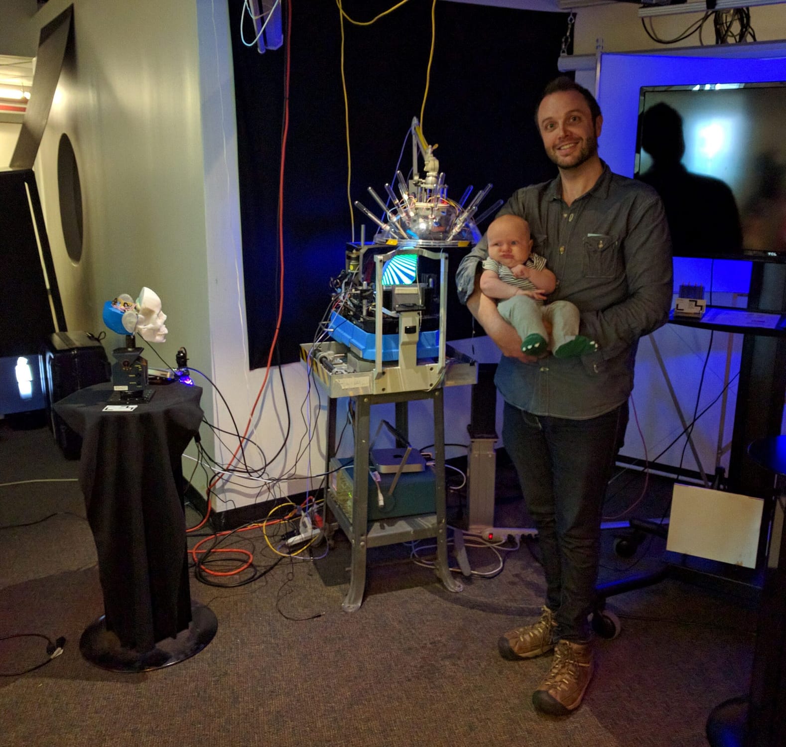
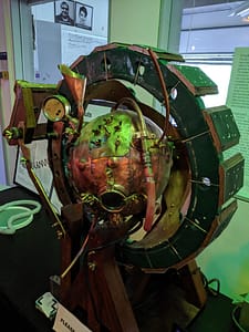
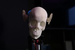
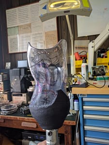
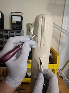
Building animatronic substrates for calibration and testing of bionic interfaces.
This invention comprises an apparatus for retinal self-imaging. Visual stimuli help the user self-align their eye with a camera. Bi-ocular coupling induces the test eye to rotate into different positions.
Model or manifest, the eye plays catalyst to the senses. For me, a gateway, separating the external
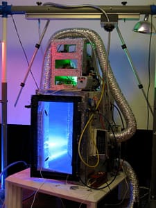
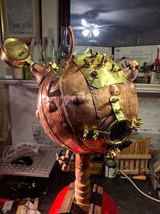
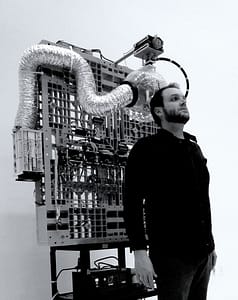
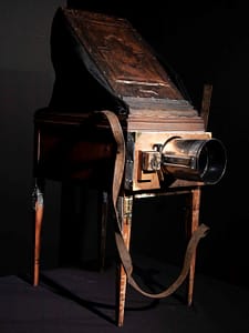
I engineered and built a 12 cubic foot vacuum particle accelerator with a power grid running at 300,000 V made from discarded television components, and a 10,000-watt stepped-up magnetron series. It is a photographic platform with which I have the freedom and flexibility to address a variety of natural mediums at the molecular level.
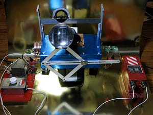
Through the use of microwave and radio transmissions, manipulated by musical compositions, I compose living organic matter orchestrated through the manipulation of frequency.
There is an inextricable link between the perspective of human engagement and the reaction of unseen forces.
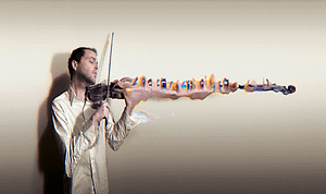
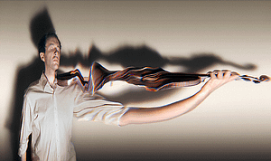
The Art of Tone is a visual approach to the granular synthesis of sight and the very nature of particles specific to our perspective in space and time
I created a number of artificial retinas on which images can be focused to address iconic decay as an unperceived aspect of sight. These carefully arranged elements have been developed into a device which can only “see” afterimages, presenting an aesthetic world of imagery beyond our conscious view.
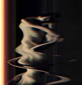
These pieces are single exposures made possible by the first digital camera I designed and built, which is nearly three feet across, makes an absolutely horrible noise, and has enough copper in it to make about 2000 pennies.
Thinking of the human visual system as a complex matrix of time-limited interactions, which are constantly decoding

Miner’s pick mattock, a differential joint from a piece of old farm equipment, a mining rail, and
ABSTRACT: A projector and one or more optical components project a light pattern that scans at least a portion of an anterior segment of an
Indirect, diffuse illumination may be employed, rather than direct illumination of the retina through the pupil. For indirect illumination, a cool-to-the-touch light source is pressed