Capturing dynamic images of the eye are a critical component to understanding the health of the visual system, but also reveal critical biomarkers for the overall well being of the entire body. This approach to retinal imaging, with the introduction of light field capture, on a micro camera platform, allows for a robust approach to the acquisition of multi-view capture ensuring nearly all light emitting from the pupil is gathered for processing. The potentials of such application in the field will provide a computational approach to processing, coupled with novel capture that may become an important non-invasive rapid screening tool for the early detection and monitoring of certain diseases, with the initial through-put of providing a rapid screening device to measure and monitor glaucoma, one of the most common causes of blindness in the world.
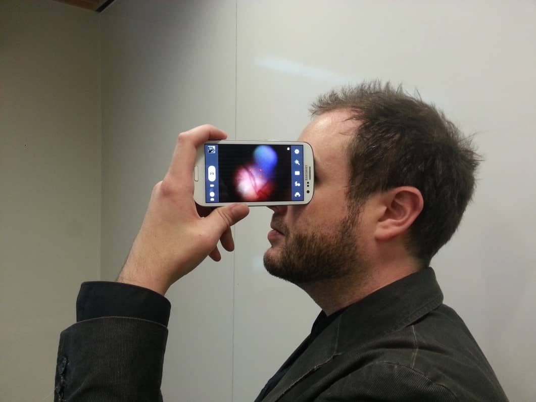
Traditionally, cell phone based fundus imaging is performed with the addition of lenses placed between the camera and subject, or as an attachment to other ophthalmic devices to achieve optimal magnification and off-axis illumination delivery. However, this requires non trivial alignment and expert knowledge to operate. The native optics of the cell phone offer the opportunity to image the retina for a novice user. Micro waveguide technology delivers specular free imaging. The hardware complexity is replaced with sophisticated software methods for non expert control and image reconstruction.
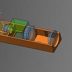
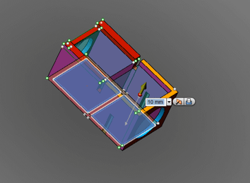
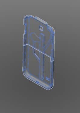
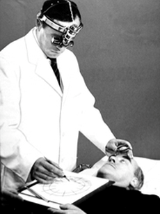
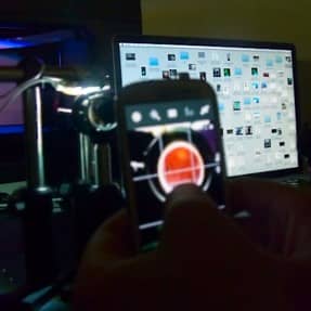
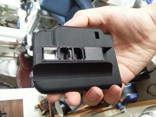
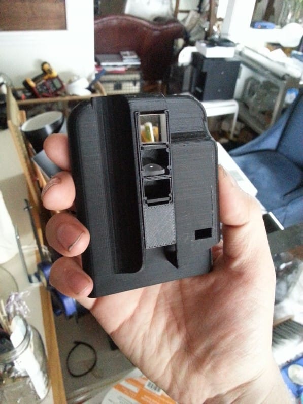
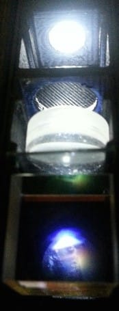
Prototype 1
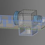
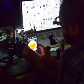
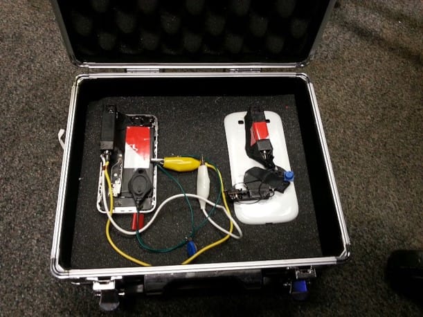
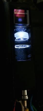
Prototype 2
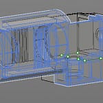
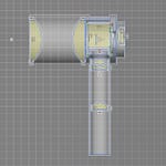
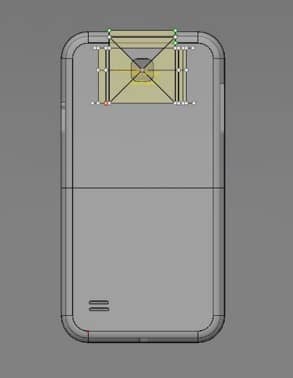
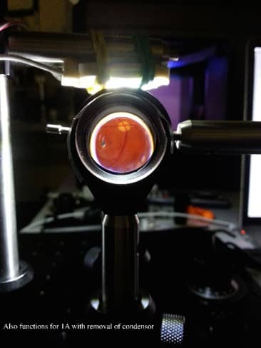
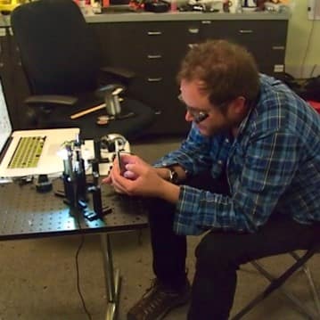
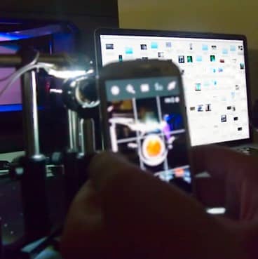
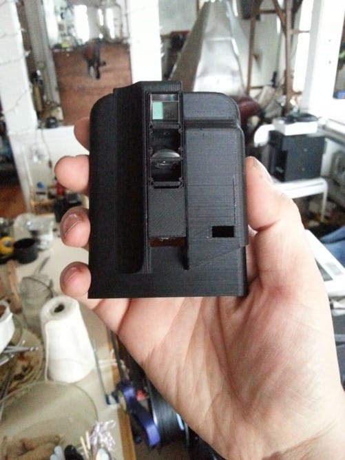
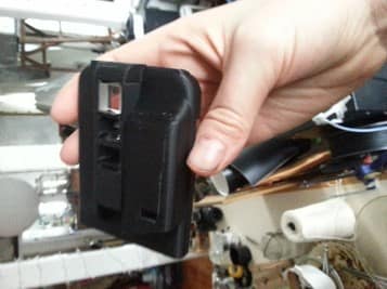
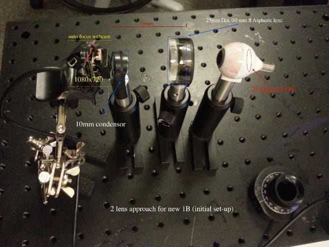
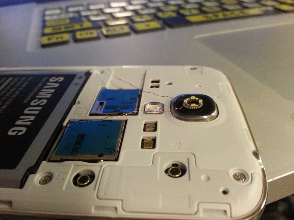
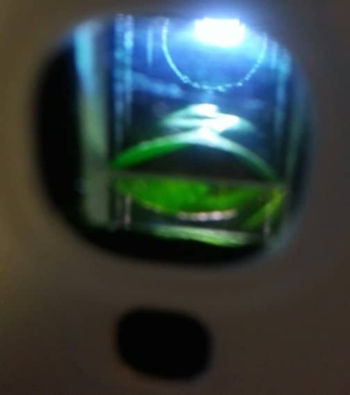
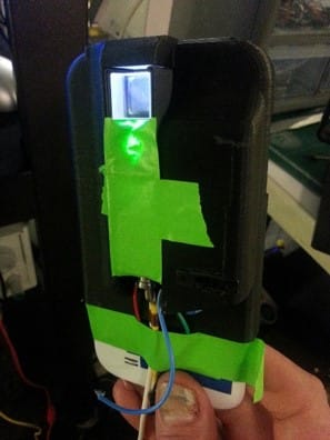
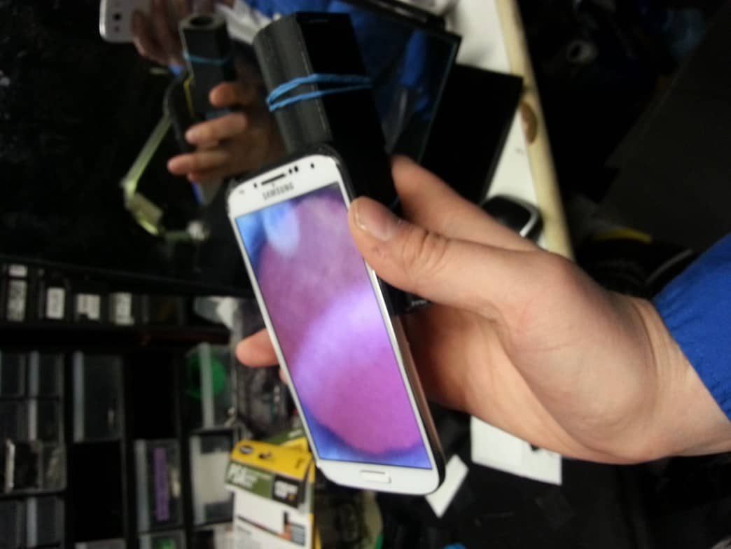
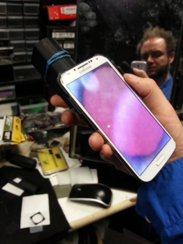
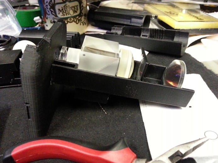
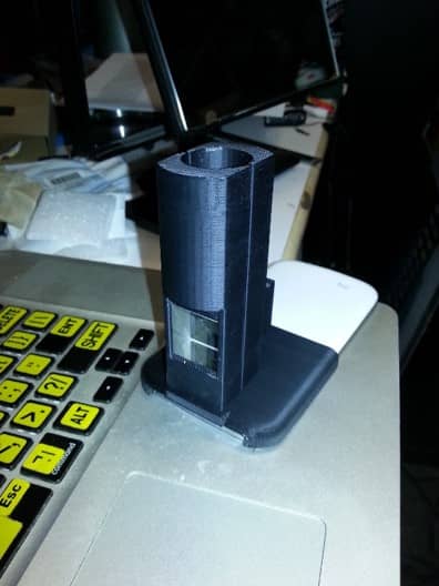
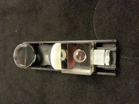
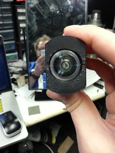
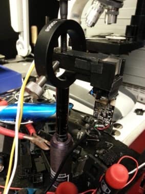
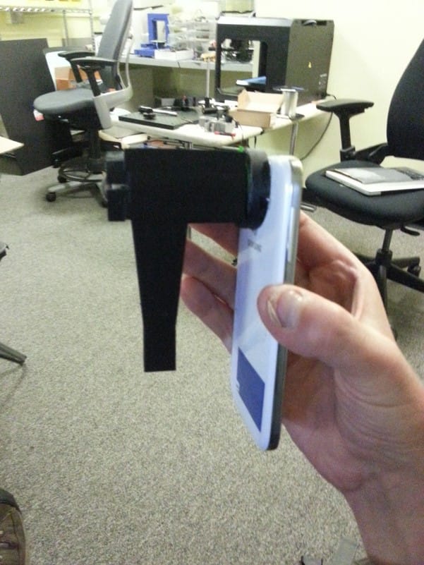
Prototype 3
Non mydriatic fundus imaging on smart phone platforms may prove to be a paradigm shift in rapid screening devices with no operational expertise required. In contrast to the standard imaging systems, the over-all data acquisition necessary for comprehensive analysis can be gathered via single capture on a micro camera configuration coupled with a computational approach.
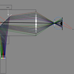
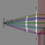
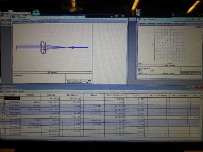
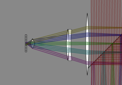
The system comprises of a smart phone (for initial studies a Galaxy S3 was chosen) coupled with a novel programmable waveguide to steer near co-axial illumination through the pupillary plane (Figure 3). The system is held 25mm from the surface of the cornea co-axial to the foveal center. The wave guide is coupled with the rear facing camera in a small clip-on element. Successive images are captured under varying computationally driven illumination strategies using a user driven visual stimulus control. A standard 500 mW retinoscope LED is applied in combination with a programmable mask and polarized beam splitter to steer a projected 2mm beam through the pupillary plane.
The S3 sensor is an 8MP (SONY) enabling image capture at 30 HZ, while the rapid auto focusing is disabled and camera parameters are controlled manually through software. The native aperture of 2.8 f-stop is used with a pixel pitch of 1.4 um.
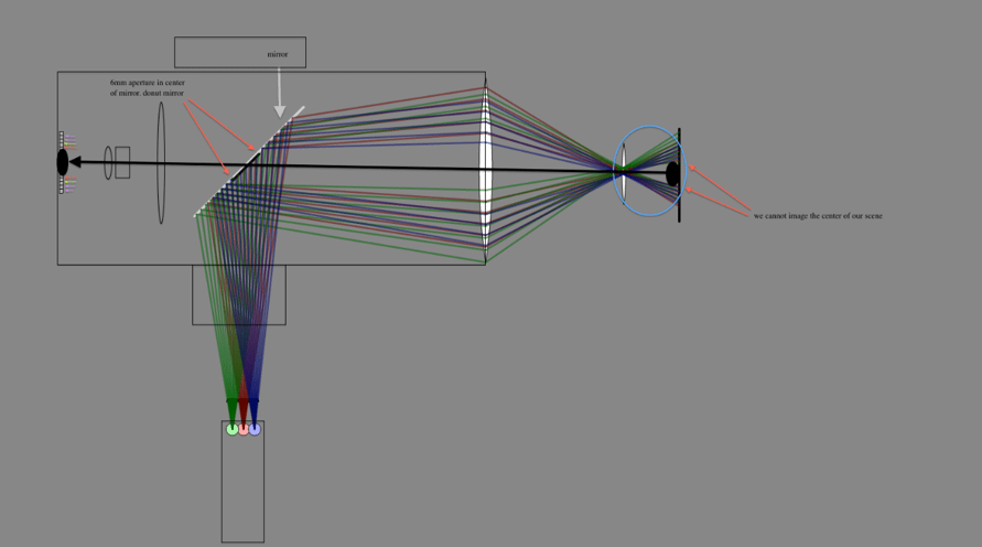
Wide-field, self-guided neurovascular imaging: additional illumination path
Results:
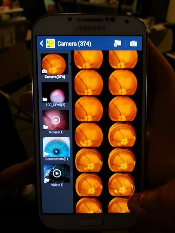
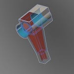
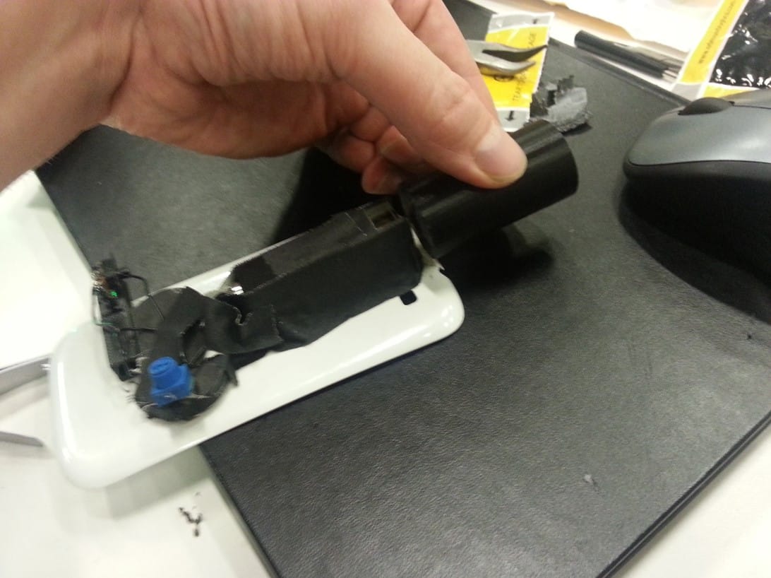
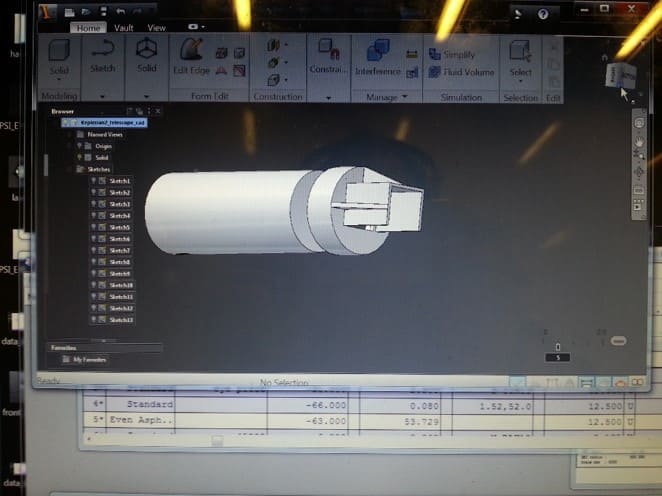
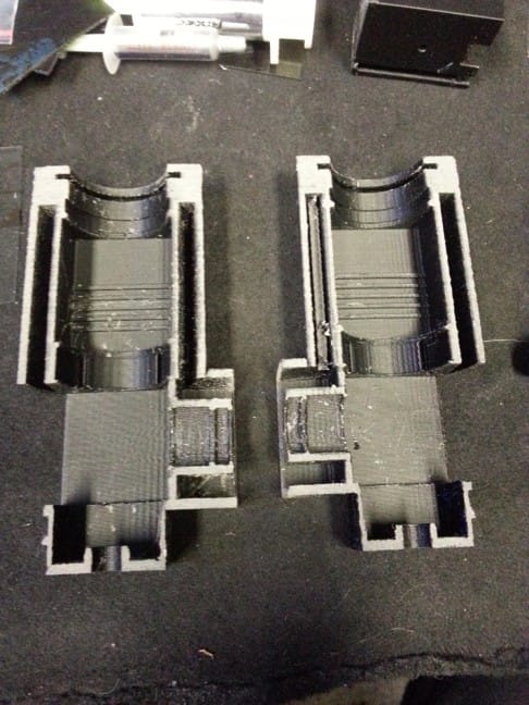
Prototyping process images:
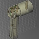
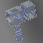
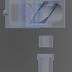
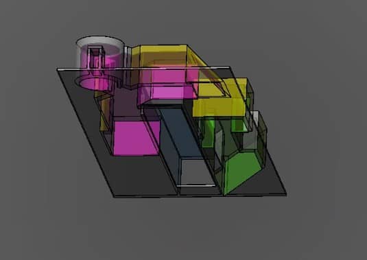
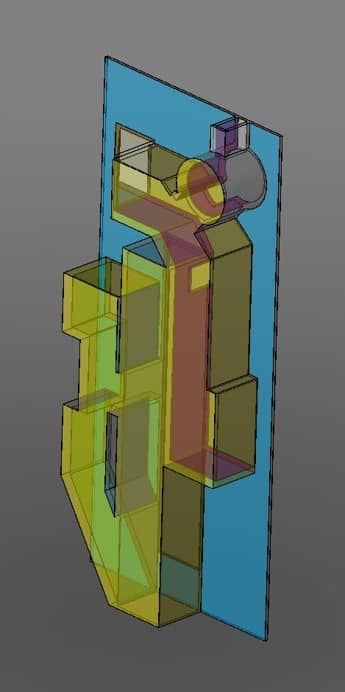
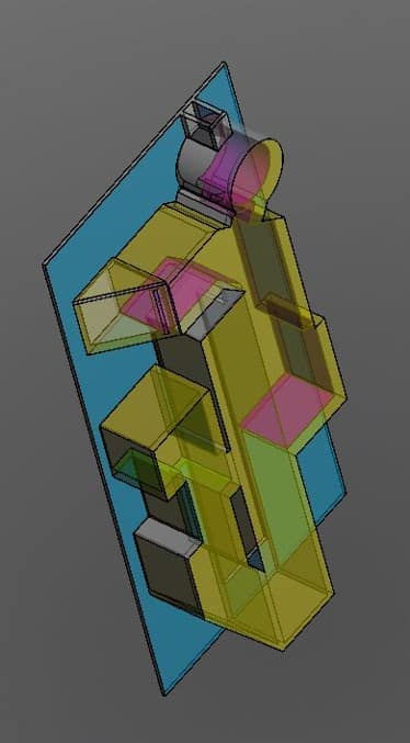
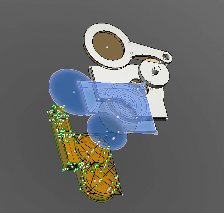
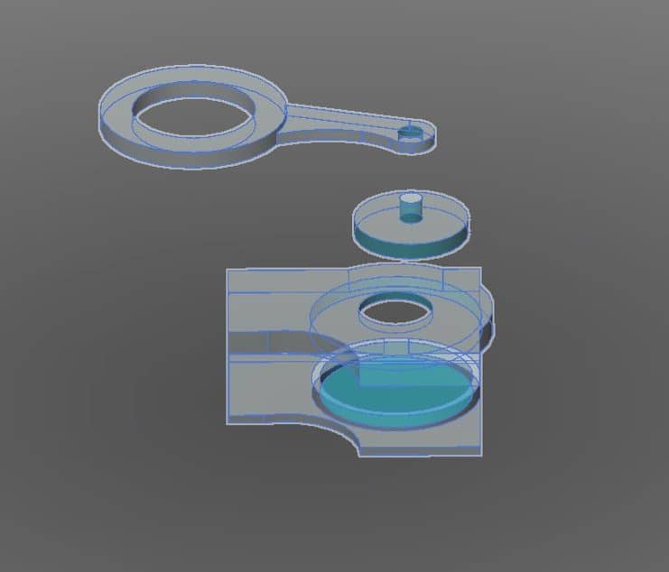
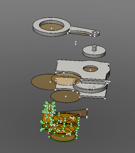
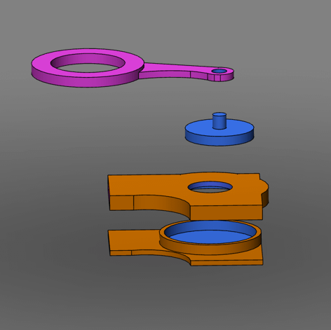
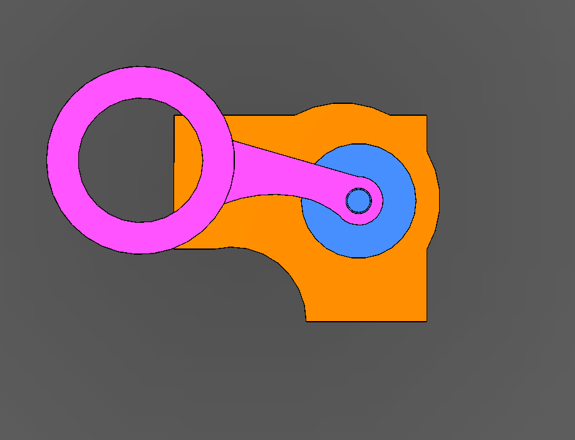
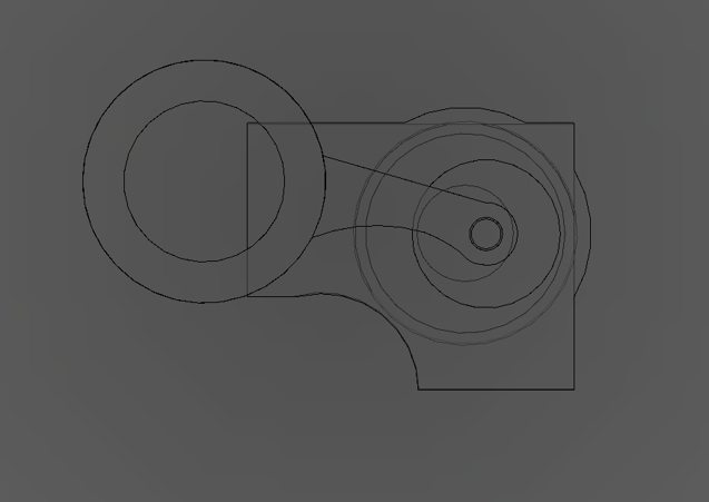
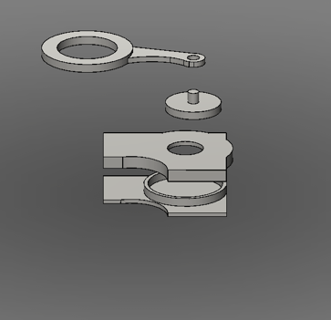
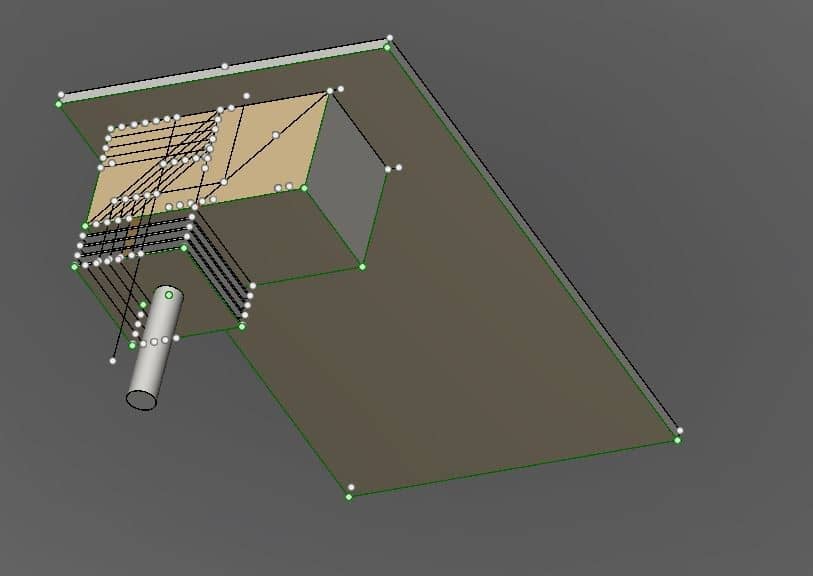
CAD design examples: