See patent Methods and apparatus for retinal imaging
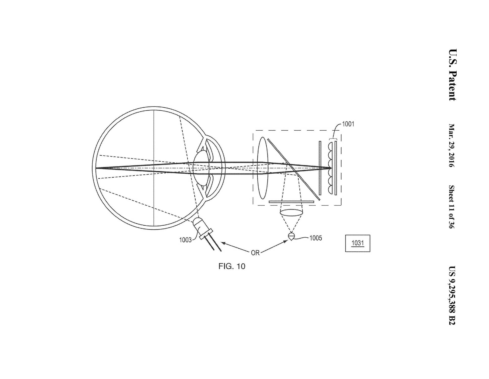
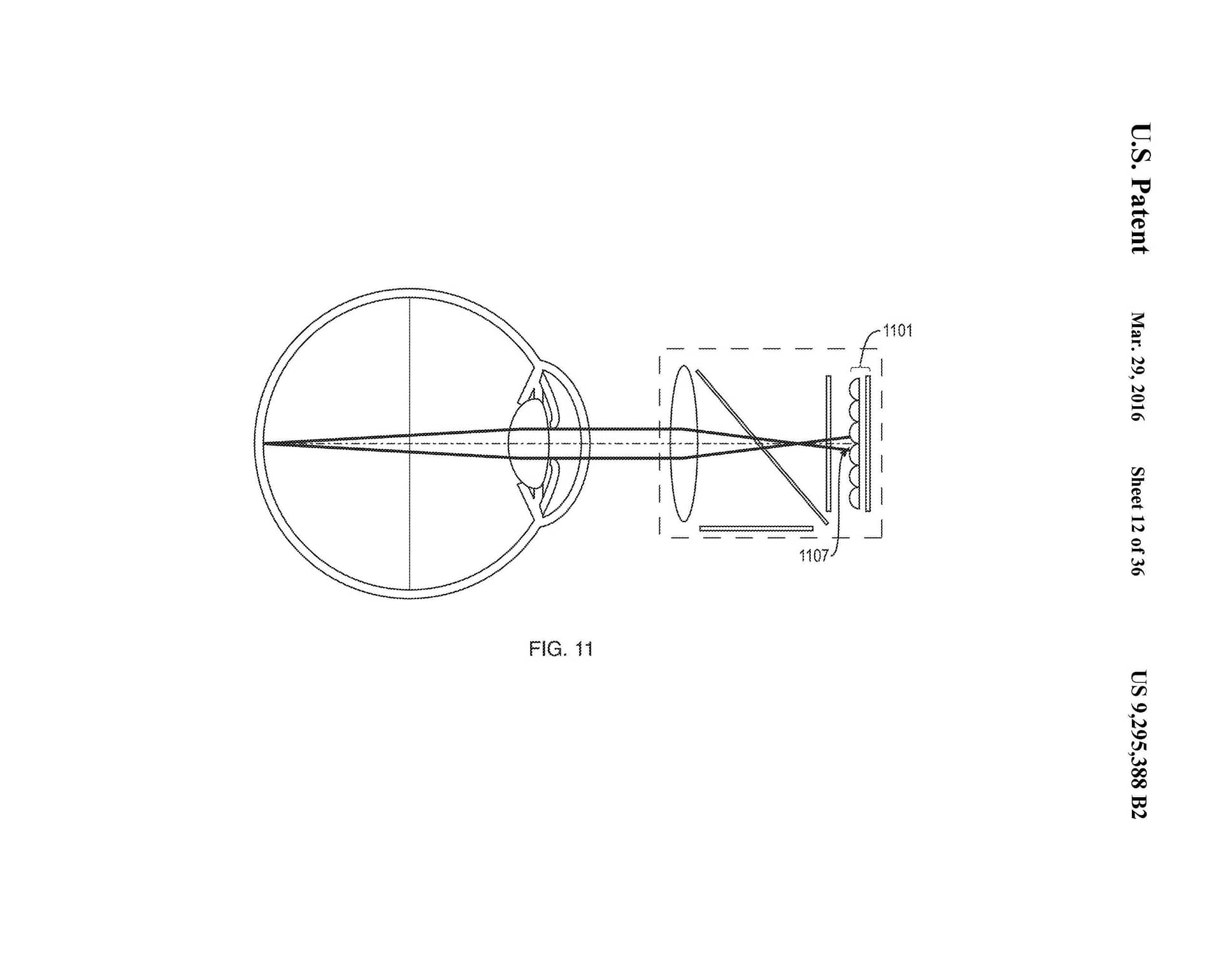
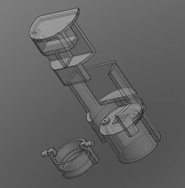
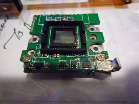
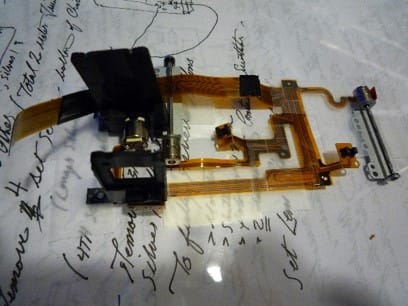
This system introduces ophthalmic light field capture, on a micro camera platform, allowing for a robust approach to the acquisition of multi-view imaging and ensuring nearly all light emitting from the pupil is gathered for processing.
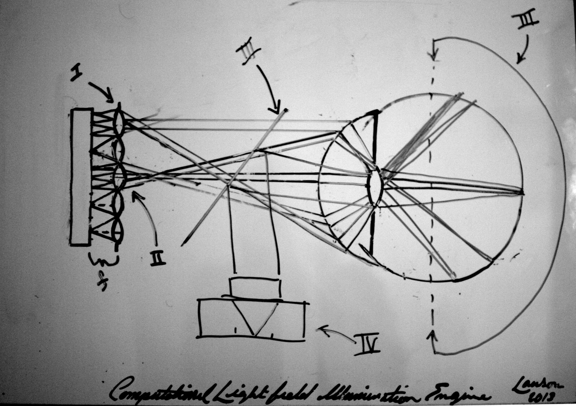
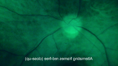
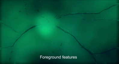
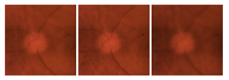
Conventional retinal modalities and instrumentation in the clinic use complex auto-focusing mechanisms to overcome the optical properties of the eye in order to achieve a narrow depth of field across the fundus, with temporally modulated triggered strobe illumination to capture a sharp image of the retina. The explorations in this prototype reduced to practice are toward a new generation of fundus imaging techniques, which utilize a 100 fold reduction in hardware by removing the need for rapid auto-focusing mechanisms or strobe illumination triggering for capture. Instead, this work explores light-field imaging to allow for re-focusing of the image after it has been taken with the addition of 3D information of the retinal architecture. In these systems the image plane is replaced by a lenslet array, re-imaging the main lens aperture. These coded rays provide directional information (light-field) onto the image plane. There is a trade off of spatial resolution for 4D light-field information, yet these are the first steps toward redefining optical engineering for ease of use in fundus photography, in addition to enabling digital refocusing with post processing techniques.
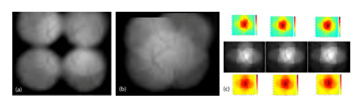
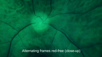
Multi-channel refocusing through retinal layers: