Novel slit lamp architecture: solid state instrumentation toward a computational platform (see patent)
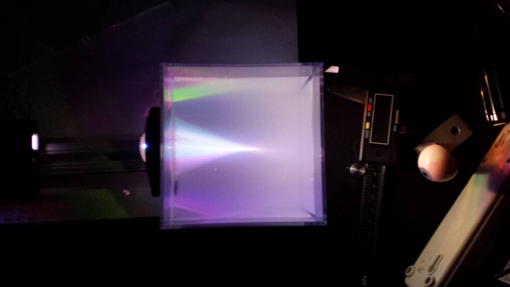
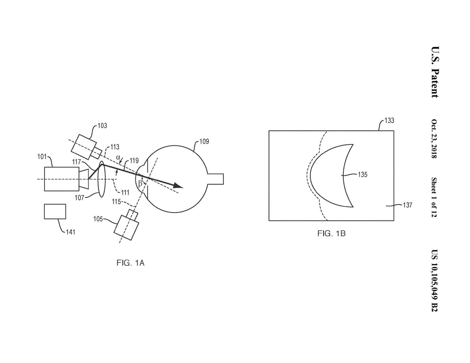
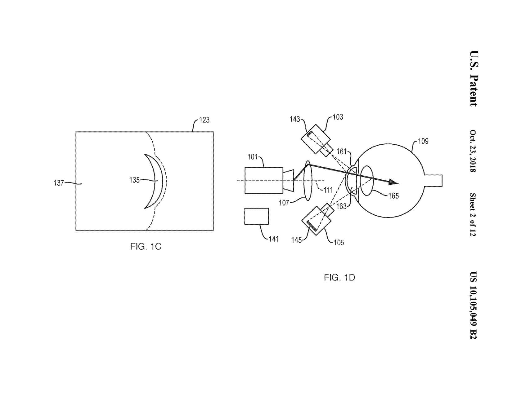
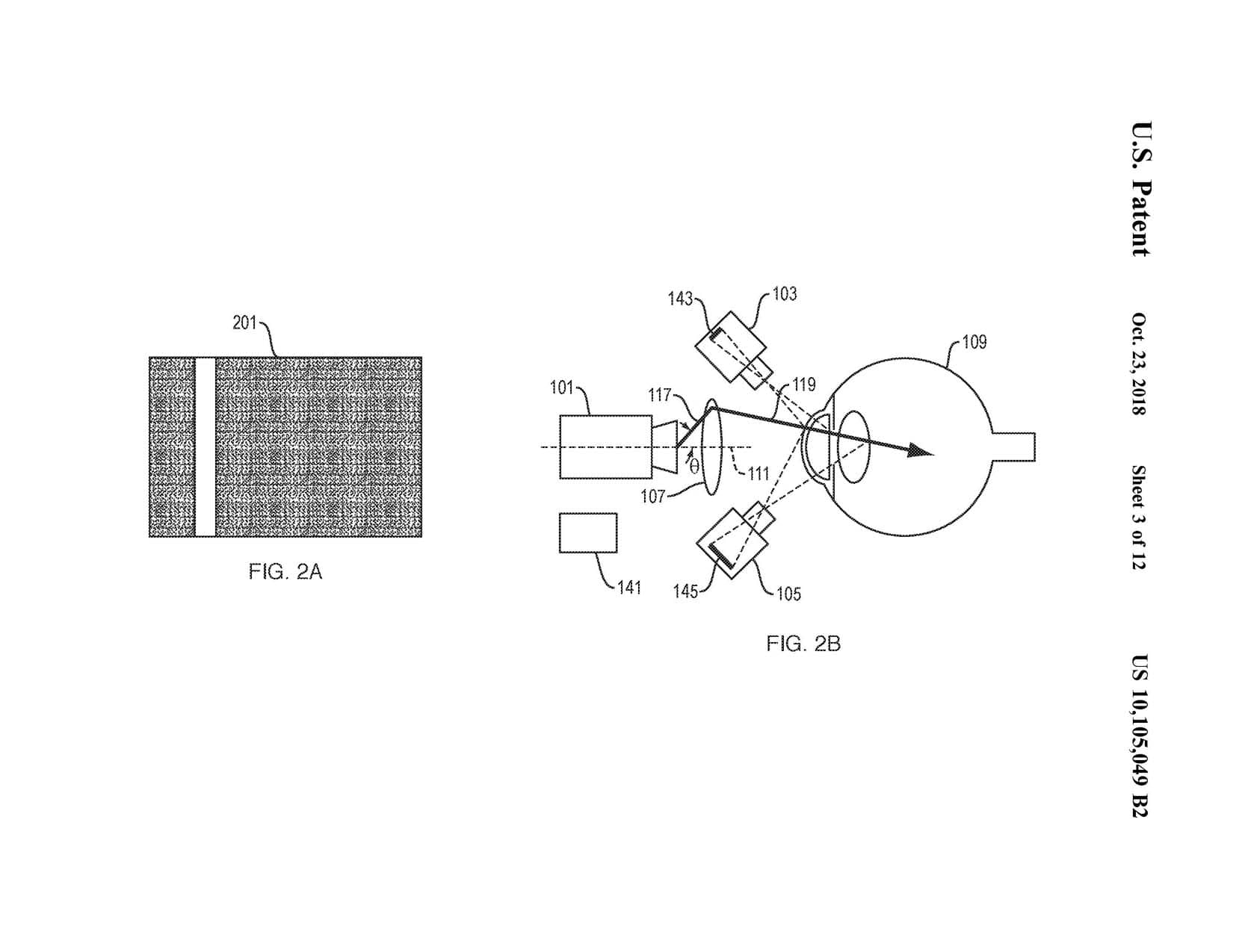
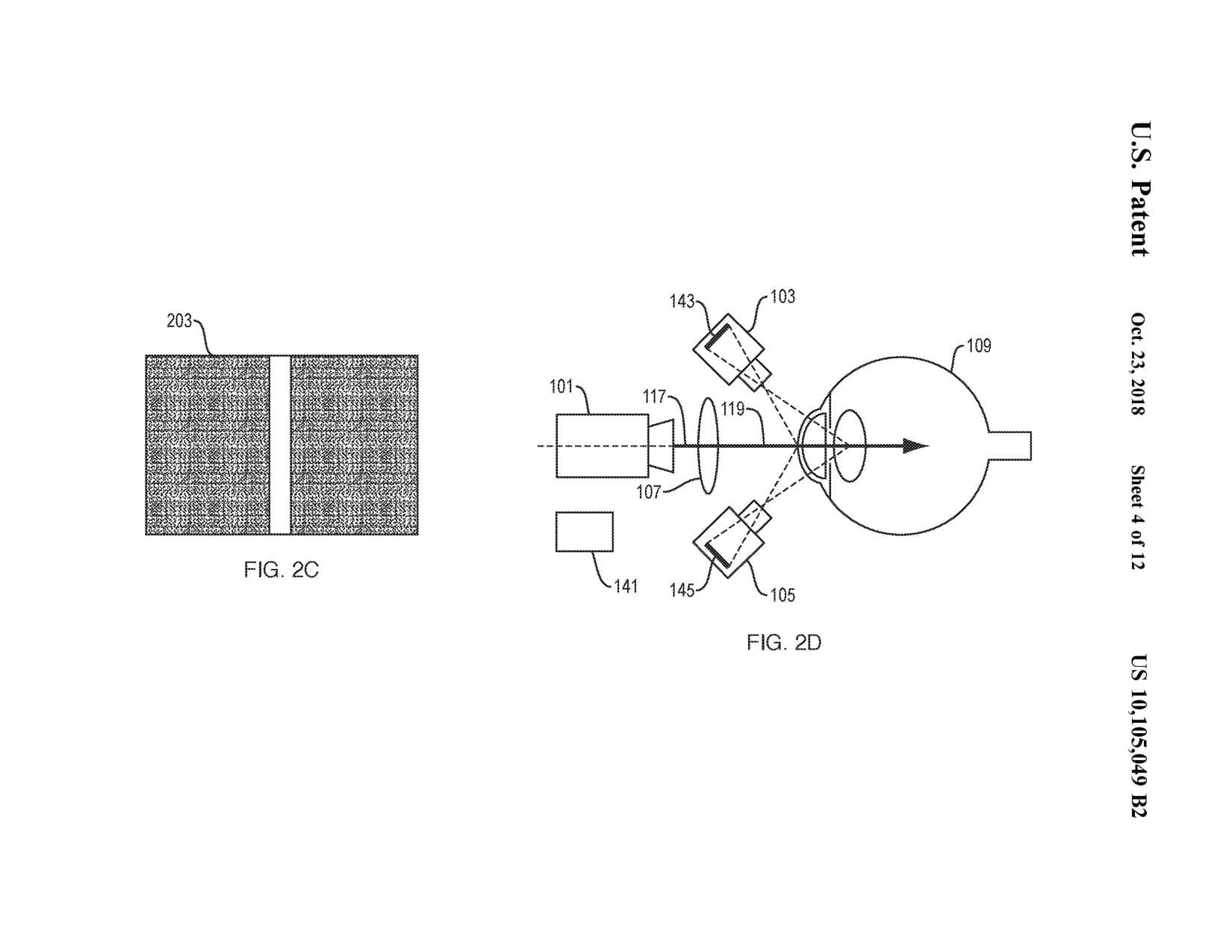
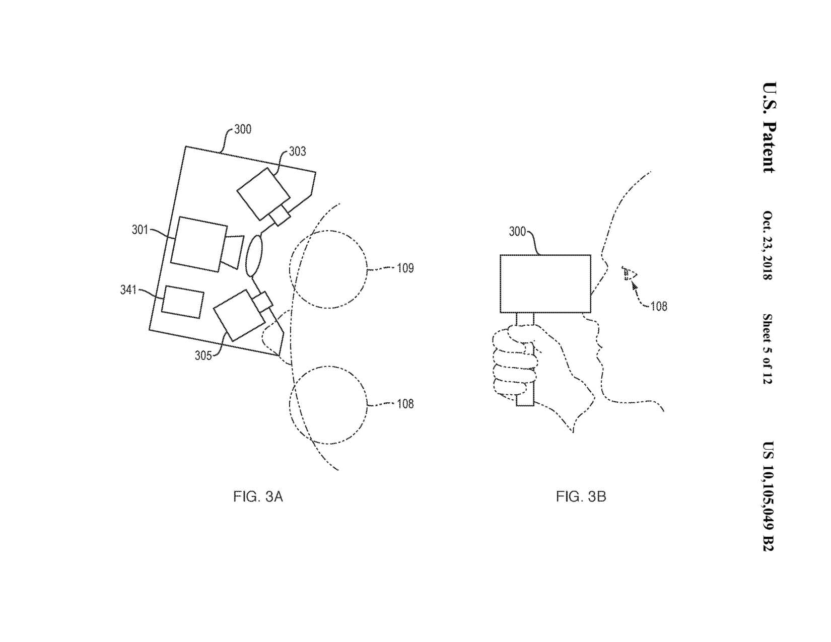
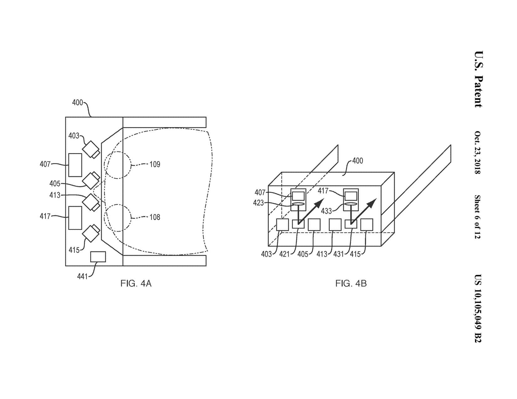
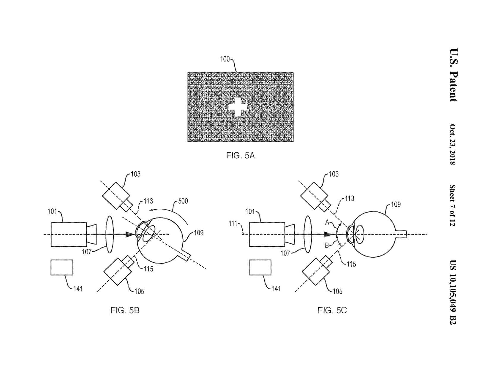
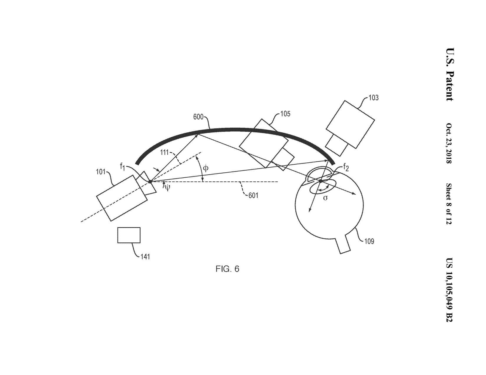
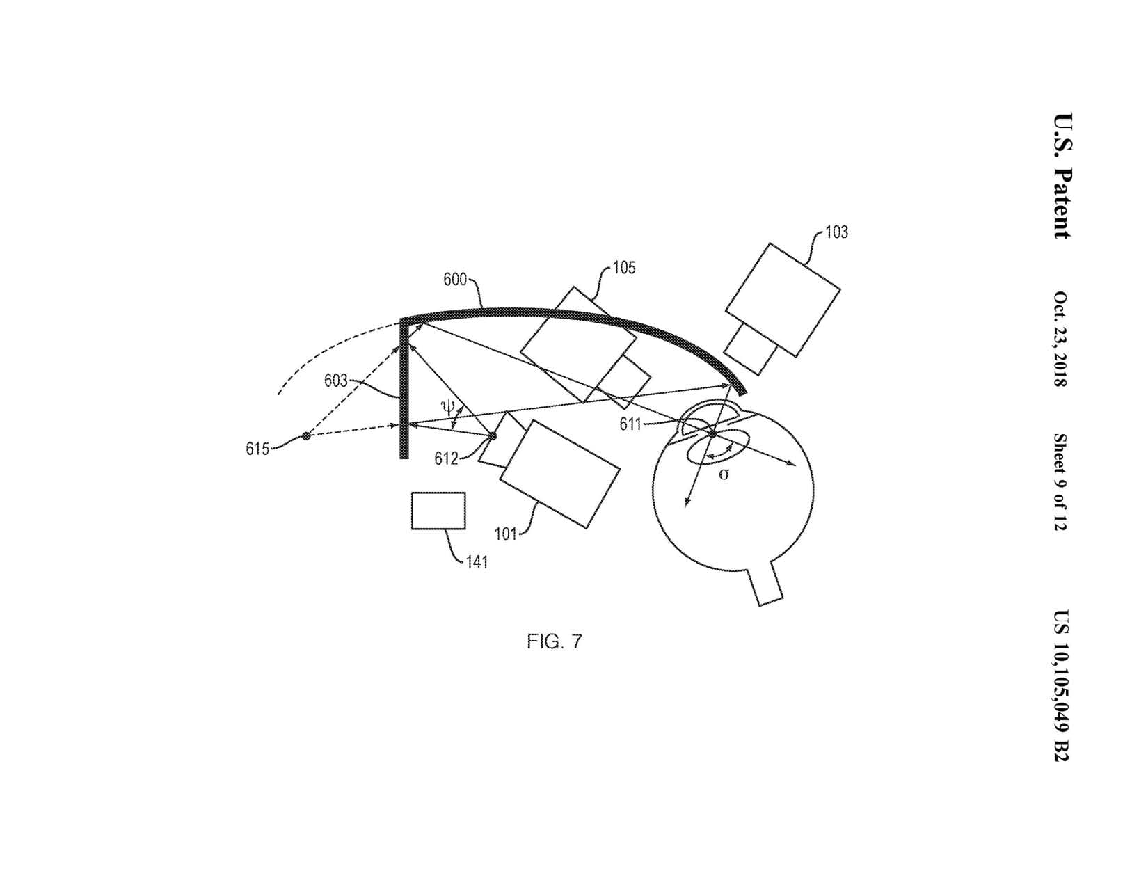
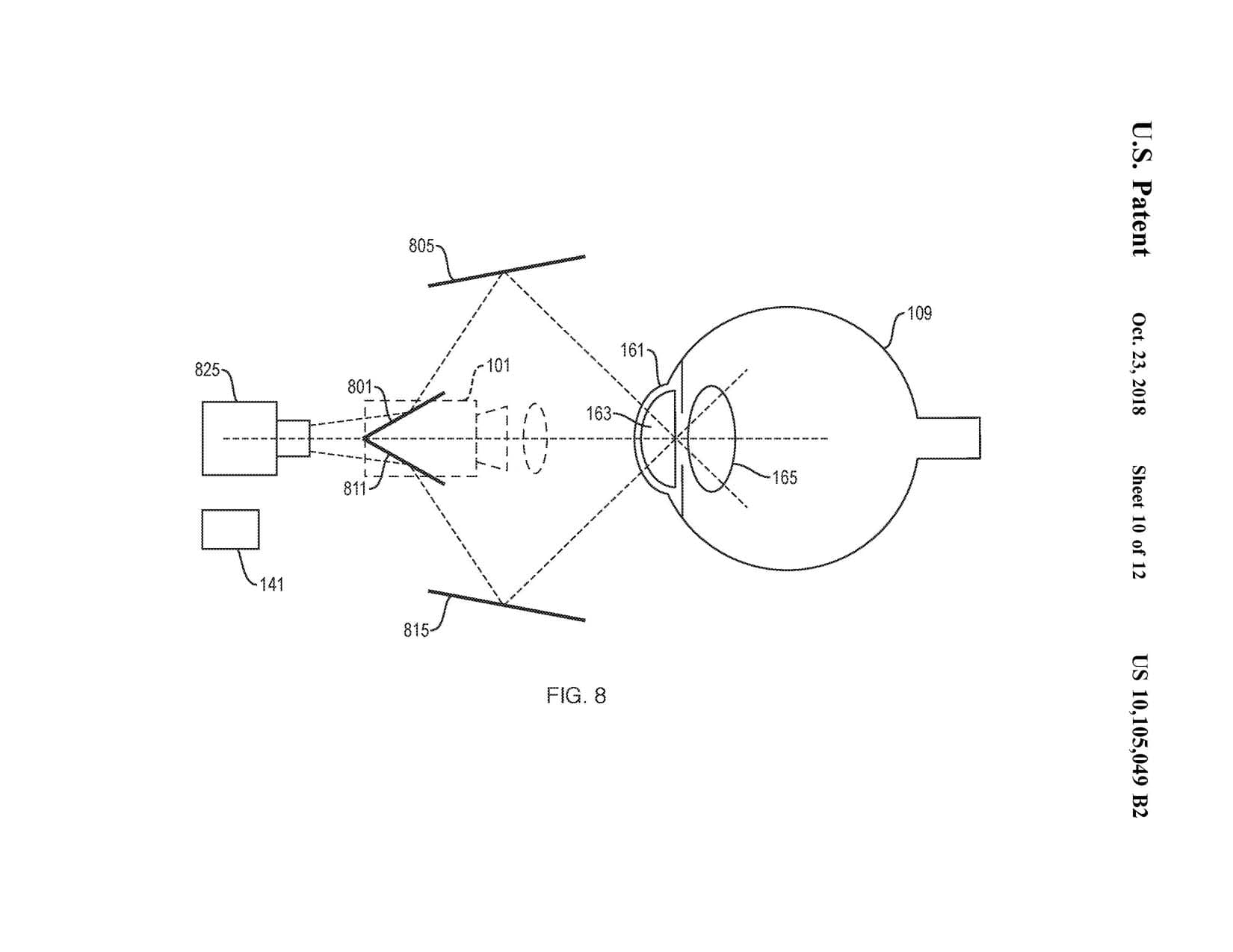
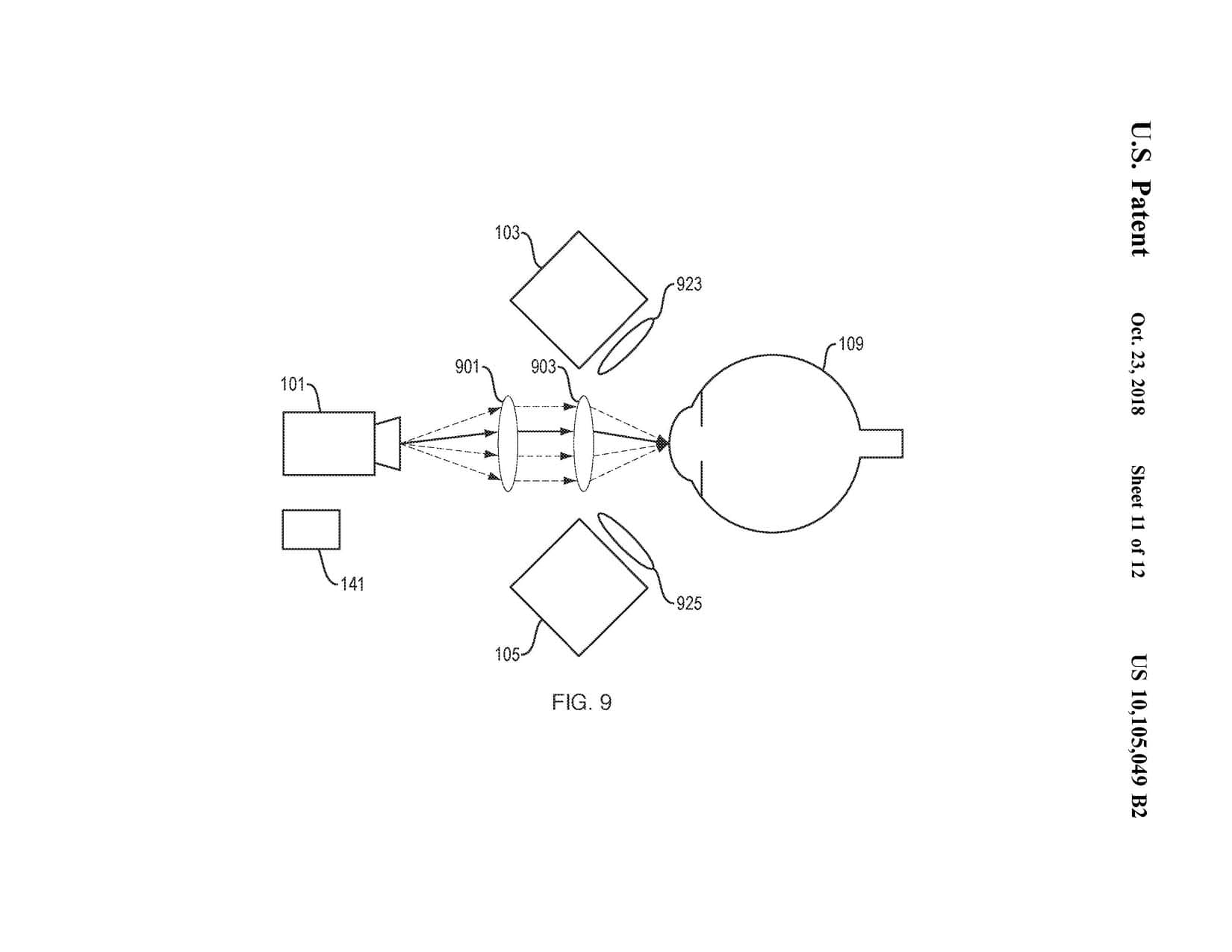
In illustrative implementations of this invention, a low-cost, wearable solid-state device with no moving optical parts is used to create a full 3D reconstruction of the anterior segment of the eye (including the corneal epithelium and endothelium, the iris, the pupil and the crystalline lens). A pico-projector projects computationally generated patterns onto the eye and one or more cameras image the scattering produced at the boundary of optical media with different refractive indices. A computer executes a software program to reconstruct a 3D model of the anterior segment of the eye. The computer uses the generated 3D model to produce topographical maps of the anterior and posterior surfaces of the cornea, measure the curvature at any point of the cornea and to measure corneal thickness.
Further anterior segment applications and results:
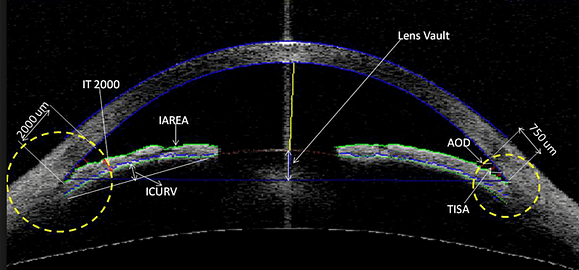
The human eye has two principal anatomical segments: the anterior segment and the posterior segment. The anterior segment includes the cornea, iris, lens, ciliary body, and the anterior portion of the sclera. It also includes the anterior chamber, which lies between the cornea and the iris, and the posterior chamber, which lies between the iris and the lens. Both the anterior and posterior chambers are filled with aqueous humor.
Many disorders, such as cataracts, ulcers, pterygia, and angle-closure glaucomas affect the anterior segment of the eye and can lead to blindness if left undiagnosed.
Conjuctiva
Tear film
Pupil
